
How to Identify a Dental Cavity: Signs, Stages & What It Looks Like
What Is a Dental Cavity?
Definition of a Dental Cavity
A dental cavity is a permanently damaged area of the tooth where the hard outer surface has broken down and formed a small opening or hole. The clinical term is dental caries, though most people simply call it a cavity or tooth decay. Once that damage occurs, it does not reverse on its own. The hole exists, and without treatment, it will grow larger over time as more of the tooth structure is eaten away.
Cavities are not just a cosmetic issue. They represent a real structural problem within the tooth, and depending on how far the decay has progressed, they can affect the enamel, the deeper layer of dentin, and eventually the soft inner pulp where nerves and blood vessels live. What starts as a tiny spot of damage can become a serious infection if left alone long enough.
How Cavities Develop on Teeth
The development of a cavity is a gradual process that typically unfolds over months or even years. It begins with the bacteria that naturally live in the mouth. These bacteria feed on the sugars and starches left behind on tooth surfaces after eating and drinking. As they digest those food particles, they produce acid as a byproduct. That acid is what causes the trouble.
Over time, repeated acid exposure weakens the mineral content of the enamel. The enamel begins to lose calcium and phosphate, two minerals essential to its hardness and integrity. This process, known as demineralization, makes the enamel increasingly vulnerable. Eventually the surface softens enough that a hole forms, and the decay process begins advancing into the deeper layers of the tooth.
The Connection Between Plaque, Bacteria, and Tooth Decay
Plaque is the sticky, colorless film that forms on teeth throughout the day. It is made up largely of bacteria, and when it is not regularly removed through brushing and flossing, it hardens into tartar, which is much more difficult to clean off. The bacteria within plaque are the direct cause of cavities because they are the ones producing the acid that erodes enamel.
The most significant cavity-causing bacterium is Streptococcus mutans, though it is not the only one involved. These organisms thrive in the warm, moist environment of the mouth, and they are particularly active after meals when sugar is available as fuel. The more frequently teeth are exposed to sugar, and the less thoroughly plaque is removed, the more opportunity these bacteria have to cause damage.
Why Cavities Are One of the Most Common Dental Problems
Dental cavities rank among the most widespread chronic health conditions globally, affecting people of every age, background, and income level. Children are frequently diagnosed with cavities in their primary and permanent teeth, but adults are far from immune. In fact, many adults develop cavities between teeth and around the edges of existing fillings that they would never notice on their own without a dental examination.
Part of the reason cavities are so common is that the conditions required for them to develop are present in virtually everyone’s mouth. Sugar consumption is nearly universal, oral bacteria are always present, and even the most dedicated brushers tend to miss certain areas consistently. Without regular professional cleanings and examinations to catch problems early, cavities have plenty of opportunity to develop and progress undetected.
What Does a Dental Cavity Look Like?
Early Signs of a Cavity on a Tooth
In the earliest stages, cavities are often invisible to the naked eye and virtually impossible for a patient to detect without professional help. The visual changes that do appear are subtle, and it takes a trained eye and proper lighting to recognize them for what they are.
White Spots on Tooth Enamel
The first visible sign of a developing cavity is often a white spot on the tooth surface. This white or chalky discoloration is a sign that the enamel in that area has begun to lose its mineral content. The spot may look slightly different in texture and sheen compared to the healthy enamel around it. At this stage, the damage is still confined to the outer surface, and with the right interventions, the process can sometimes be slowed or reversed before a true hole forms.
Chalky or Rough Tooth Surfaces
Healthy enamel has a smooth, slightly glossy surface. When demineralization is underway, the affected area can feel rough or chalky when you run your tongue across it. This textural change reflects the breakdown of the enamel’s crystalline structure. It is easy to miss, particularly on back teeth, but it is a meaningful early warning sign that something is happening beneath the surface.
Moderate Cavity Appearance
As the cavity progresses past the early enamel stage and begins to move deeper into the tooth, the visual changes become more pronounced. At this point, a careful look in a well-lit mirror might actually reveal something concerning.
Brown or Dark Stains on Teeth
A cavity that has progressed beyond the initial white spot stage often begins to appear as a tan, brown, or dark discoloration on the tooth. This happens because the decayed area absorbs pigments from food, drinks, and bacteria over time. The color can range from light yellowish brown to nearly black depending on how long the decay has been present and how deep it has gone. It is worth noting that not every dark spot is a cavity, but any discoloration that is new, growing, or sitting in a groove or pit deserves professional attention.
Visible Pits and Grooves
As the decay eats through more of the enamel, it begins to create small pits or deepening grooves in the tooth surface. These are most noticeable on the chewing surfaces of back teeth where the natural anatomy already features fissures and valleys. A cavity can make these normal grooves noticeably deeper or can create entirely new depressions in the enamel that were not there before. Food tends to collect in these areas, which accelerates the decay process even further.
Advanced Cavity Appearance
When a cavity has been present and untreated for a significant period of time, the visual damage becomes much harder to ignore. At this stage, the tooth structure itself has been substantially compromised.
Holes in the Tooth Surface
A cavity that has been left to progress will eventually create a visible hole in the tooth. This is what most people picture when they think of a cavity, even though the damage was well underway long before it reached this point. These holes can vary in size from small perforations to large craters depending on how advanced the decay is and where on the tooth it is located.
Large Areas of Decay
In some cases, particularly when treatment has been delayed for an extended period, the decay can spread across a large portion of the tooth surface. What begins as a small pit can eventually involve multiple cusps or surfaces of the same tooth. This level of damage often requires more extensive restorative work than a simple filling can address.
Broken or Crumbling Teeth
When decay has been present long enough to undermine a significant portion of the tooth’s internal structure, the remaining enamel and dentin can become fragile and susceptible to fracture. Patients sometimes notice that a tooth feels rough, sharp, or different when touched with the tongue, or they may discover that a piece of the tooth has actually broken off. This usually occurs without any dramatic incident, sometimes just from normal chewing on a tooth that has been weakened from within.
Cavity Appearance on Different Types of Teeth
Front Teeth
Cavities on the front teeth are often the easiest for patients to notice on their own, simply because they are visible when you smile or open your mouth. They tend to develop on the surfaces facing the lips or, more commonly, between adjacent front teeth where floss would normally clean. Early cavities between front teeth can appear as small white spots along the gumline or as faint discoloration between the teeth that becomes more obvious as the decay advances.
Molars and Premolars
The back teeth are the most cavity-prone in the mouth because their wide, grooved chewing surfaces create ideal hiding places for bacteria and food debris. Cavities in the pits and fissures of molars are extremely common and often progress significantly before causing noticeable symptoms. They may appear as dark staining in the grooves that looks different from ordinary surface discoloration, or as areas where the tooth surface seems to sink inward when examined closely.
Between Teeth
Interproximal cavities, meaning those that form between adjacent teeth, are among the most difficult for patients to see on their own and among the most common types that dentists find on X-rays. There is no visible opening to spot in the mirror because the decay is hidden in the tight contact zone between two teeth. By the time a cavity between teeth becomes large enough to notice visually, it has usually already caused significant damage to both of the teeth it sits between.
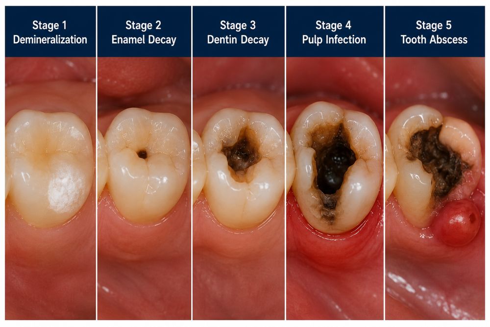
Understanding the Stages of a Dental Cavity
Stage 1 – Demineralization
Loss of Minerals in Tooth Enamel
Demineralization is the very beginning of the cavity process, and at this point no true cavity has formed yet. The enamel is losing minerals faster than it is able to replace them through natural remineralization, which occurs when saliva delivers calcium and phosphate back to the tooth surface. When the balance tips too far toward mineral loss, the enamel becomes weakened and vulnerable to further damage. This stage is reversible under the right conditions, which makes it the most important time to intervene.
Early White Spot Lesions
The outward sign of active demineralization is the white spot lesion. These opaque white or chalky patches on the enamel surface represent areas where the mineral density has dropped significantly. A dentist or hygienist trained to look for them can identify white spot lesions during a routine examination, though they are easy to overlook and not visible on traditional dental X-rays. Fluoride treatments, remineralizing agents, and improvements in oral hygiene can sometimes stop the progression and help the enamel partially recover at this stage.
Stage 2 – Enamel Decay
Small Surface Cavities
When demineralization is not caught and the enamel continues to weaken, the surface eventually breaks down and a small cavity forms. At this point the damage is still confined to the enamel and has not yet reached the dentin below. These early surface cavities are typically treated with a small composite filling and require minimal removal of tooth structure. The sooner they are caught, the simpler and less expensive the repair tends to be.
Increased Sensitivity
Some patients begin to notice sensitivity during this stage, particularly to sweet foods or cold temperatures. The enamel, which normally insulates the tooth, has been compromised enough that certain stimuli are able to reach the nerve fibers more easily. Not everyone experiences this sensitivity, however, and its absence does not mean a cavity is not present. Many people have no symptoms at all until the decay has progressed to a more advanced stage.
Stage 3 – Dentin Decay
Deeper Tooth Damage
Dentin is the layer of tooth structure that lies just beneath the enamel. It is softer than enamel and contains tiny fluid-filled channels called dentinal tubules that connect to the nerve of the tooth. Once decay has broken through the enamel and entered the dentin, it progresses more quickly because dentin is less resistant to the acids that bacteria produce. A cavity at this stage requires a filling, and depending on how much dentin has been affected, a larger restoration may be necessary.
More Noticeable Symptoms
Because of the dentinal tubules, decay in the dentin layer tends to produce more obvious symptoms than enamel-only cavities. Sensitivity to temperature and sweetness becomes more pronounced, and some patients begin to experience brief flashes of pain or a lingering ache in the tooth. The pain may appear spontaneously or consistently in response to specific triggers. At this point, the cavity is unlikely to be invisible on a dental X-ray, and most patients will have it confirmed at their next examination.
Stage 4 – Pulp Infection
Severe Tooth Pain
The pulp is the innermost part of the tooth, containing nerves, blood vessels, and connective tissue. When decay reaches the pulp, inflammation and infection set in rapidly. The pressure from that infection within the confined space of the pulp chamber causes significant and often severe pain. This pain can be constant, throbbing, and intense enough to disrupt sleep. At this stage, a simple filling is no longer sufficient. Root canal therapy is needed to remove the infected pulp tissue, clean and seal the canals, and save the tooth.
Risk of Dental Abscess
Pulp infection creates ideal conditions for the development of a dental abscess, which is a pocket of pus that forms at the root of the tooth or in the surrounding gum tissue as the body tries to contain the spreading infection. An abscess is a serious development that requires prompt treatment, and it does not resolve on its own. If left untreated, the infection can spread to the jaw, neck, or beyond.
Stage 5 – Tooth Abscess
Signs of Advanced Infection
A dental abscess typically announces itself with persistent, throbbing pain that may radiate to the jaw, ear, or neck. The gum near the affected tooth may be swollen and tender, and a small pimple-like bump on the gum, sometimes called a fistula, can appear as the infection finds a pathway to drain. Fever, swollen lymph nodes under the jaw, and a general feeling of illness are signs that the infection is more than localized and requires urgent attention.
When Emergency Dental Care Is Needed
Any abscess that is accompanied by difficulty swallowing or breathing, significant facial swelling, fever, or a feeling of being systemically unwell requires emergency care immediately. An untreated dental infection that spreads to the spaces of the neck or the airway can be life-threatening. This is not an exaggeration. Patients who have reached this stage should not wait for a regular appointment. They should seek emergency dental or medical care the same day.
Common Signs and Symptoms of a Dental Cavity
Tooth Sensitivity to Hot, Cold, and Sweet Foods
Sensitivity is one of the most frequently reported early symptoms of a cavity. It often presents as a brief, sharp sensation when eating or drinking something cold, sweet, or acidic. Hot sensitivity tends to develop a bit later, typically when the decay has moved closer to the pulp. The sensation usually fades within seconds, which leads many people to dismiss it as normal variation. If it happens repeatedly in the same tooth or begins to intensify, it deserves professional evaluation.
Persistent Toothache or Discomfort
A toothache that lingers, returns regularly, or is becoming more intense over time is a reliable signal that something is wrong inside the tooth. Unlike sensitivity that resolves quickly, a persistent ache that lasts minutes or longer suggests the nerve is involved to some degree. Some patients describe it as a dull pressure, others as a sharp or throbbing sensation. Regardless of how it manifests, pain that keeps returning in the same location should be evaluated promptly.
Pain When Biting or Chewing
Discomfort that appears specifically when biting down or chewing can indicate a cavity that has progressed far enough to affect the structural integrity of the tooth. It can also indicate a cracked tooth or a loose filling, so a professional examination is needed to determine the cause. In any case, pain with chewing is not something to push through or ignore, as continuing to stress the affected tooth can lead to additional damage.
Visible Holes or Pits in Teeth
When a cavity has progressed to the point of creating a visible hole or noticeable pit in the tooth surface, it is well past the early stage. Patients sometimes discover these through feel rather than sight, noticing an unexpected rough edge or void when they run their tongue across a tooth. Finding a hole in a tooth is a clear sign that dental treatment is needed without delay.
Tooth Discoloration and Staining
Discoloration associated with tooth decay looks different from typical surface staining from coffee, tea, or tobacco. Decay-related discoloration tends to sit within the tooth surface rather than on top of it, and it may appear in areas that are not normally prone to staining, such as the grooves of a molar or the sides of a front tooth. Colors range from dull white or yellow to brown and black depending on the stage of decay.
Bad Breath Caused by Tooth Decay
Persistent bad breath that does not resolve with brushing or mouthwash can sometimes be traced to tooth decay. As bacteria break down tooth structure and organic matter collects in cavities, the byproducts include sulfur compounds that produce an unpleasant odor. If bad breath has become a consistent concern despite good oral hygiene habits, a dental examination to rule out decay or gum disease is a reasonable next step.
Swelling Around the Affected Tooth
Swelling of the gum tissue near a tooth, whether subtle puffiness or more pronounced inflammation, can indicate that the decay has progressed to the point of affecting the surrounding structures. In the context of advanced cavity or abscess, this swelling may be tender to the touch and accompanied by pain when biting. Swelling that appears suddenly, grows quickly, or extends into the face or neck requires urgent care.

What Does an Early Cavity Feel Like?
Mild Sensitivity Without Pain
Early cavities very often produce no significant pain. What patients might notice, if they notice anything at all, is a mild fleeting sensitivity when consuming something cold, sweet, or acidic. It does not linger, it does not wake anyone up at night, and it does not interfere with eating. Because of this, it is easy to rationalize away as something minor. But that brief sensitivity is the tooth signaling that something has changed, and it is worth paying attention to.
Temporary Discomfort After Eating Sweets
A tooth with an early cavity may react to sugary foods with a brief twinge of discomfort that disappears within moments after the food is swallowed or the mouth is rinsed. The sugar temporarily lowers the pH around the tooth, which aggravates the already-compromised enamel. This type of sensitivity specifically triggered by sweets is a pattern worth mentioning to your dentist, particularly if it seems to be localized to one area of the mouth.
Why Early Cavities Often Go Unnoticed
The unfortunate reality is that early cavities are largely asymptomatic. The enamel does not have its own nerve supply, so as long as the decay stays within the enamel layer, the tooth does not send pain signals to the brain. Patients have no way of knowing the decay is there without professional examination and imaging. This is the core reason why routine dental checkups are so valuable. A dentist examining teeth twice a year can catch cavities at a stage when treatment is simple, quick, and far less costly than waiting until symptoms appear.
Cavities in Different Areas of the Mouth
Smooth Surface Cavities
Smooth surface cavities develop on the flat outer or inner surfaces of teeth, including the cheek-facing and tongue-facing sides. These are the most preventable type of cavity because they form in areas that are fully accessible to a toothbrush. They tend to develop slowly compared to other types and are more common in people who have inconsistent oral hygiene routines. Fluoride and diligent brushing are highly effective at preventing and even reversing early smooth surface decay.
Pit and Fissure Cavities
The chewing surfaces of molars and premolars are covered in pits and fissures, which are narrow grooves and valleys that make these teeth effective for grinding food. These same grooves, however, are difficult to clean thoroughly because toothbrush bristles cannot always reach all the way in. Bacteria and food debris accumulate there easily, and cavities in pits and fissures are among the most common types seen in both children and adults. Dental sealants applied to the chewing surfaces of back teeth are specifically designed to protect these areas.
Root Cavities
Root cavities develop on the surface of the tooth root, below the gumline. They occur most often in adults and older patients whose gums have receded due to age or gum disease, exposing the root surface. Unlike enamel, the root surface is covered in a softer material called cementum, which is significantly more vulnerable to acid erosion. Root cavities can progress quickly and are often difficult to treat because of their location near or beneath the gumline.
Interproximal Cavities (Between Teeth)
Interproximal, or proximal, cavities form on the surfaces where adjacent teeth touch. They are the type most consistently missed by patients on their own because there is nothing to see from the outside. They are also the most common type caught on bitewing X-rays during routine dental visits. By the time an interproximal cavity has become large enough to cause symptoms or visible damage, it has often affected the adjacent tooth as well. Regular flossing is the primary preventive measure for this type of cavity.
Recurrent Cavities Around Existing Fillings
Recurrent decay, sometimes called secondary caries, develops at the margins of existing restorations such as fillings or crowns. Over time, the seal between a filling and the natural tooth can develop microscopic gaps where bacteria can enter and begin working on the tooth structure underneath. This type of cavity is not always apparent during a visual exam and may require X-rays to detect. It is one of the reasons dentists monitor the condition of existing restorations at every checkup.
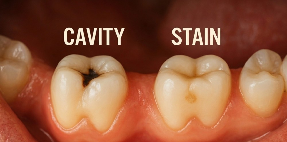
Cavity vs. Stain: How to Tell the Difference
Characteristics of Surface Stains
Surface stains are discolorations that sit on top of the enamel rather than within or beneath it. They are caused by pigments from food, beverages, tobacco, or certain medications that adhere to the outer surface of the tooth. Coffee, tea, red wine, and berries are frequent contributors. Surface stains tend to spread somewhat evenly across the enamel surface and can often be partially removed through professional polishing. They do not typically have a soft, sticky, or pitted quality to them, and they do not cause sensitivity or pain.
Characteristics of Tooth Decay
Decay-related discoloration looks and behaves differently from a surface stain. It tends to be localized to a specific spot, groove, or margin rather than spread across the whole tooth. The area may appear dull or opaque rather than simply darker, and if you were to probe it gently, the surface texture would likely feel soft or tacky rather than smooth and hard. Decay may also be accompanied by sensitivity, pain, or a rough edge that was not there before. Most importantly, unlike a surface stain, decay cannot be polished away and will continue to worsen without treatment.
When Dark Spots Require a Dental Examination
Any new dark spot, particularly one that sits in a groove, near the gumline, or between teeth, deserves professional evaluation. Even if the spot turns out to be a stain rather than decay, ruling out a cavity provides important peace of mind and ensures that any actual decay is caught early. Dentists use probes, lighting, and X-rays to determine whether a dark area represents surface discoloration or active tooth decay, and they can provide a definitive answer that no amount of at-home examination can replicate.
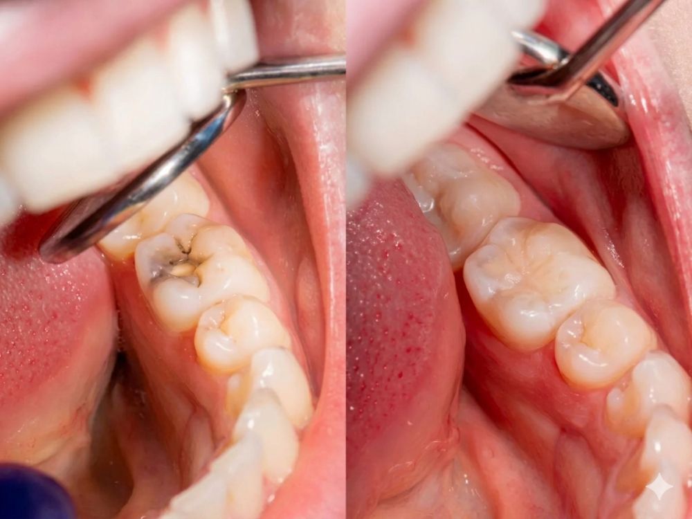
How Dentists Diagnose a Dental Cavity
Visual Dental Examination
A thorough visual examination is the foundation of cavity detection. The dentist uses a bright light, a small mirror, and a dental probe to systematically inspect every surface of every tooth. They are looking for changes in color, texture, translucency, and contour that could indicate decay. The probe helps assess whether a suspicious area on the enamel surface is hard and intact or soft and tacky, which is a reliable distinguisher between a stain and actual decay.
Dental X-Rays for Hidden Cavities
X-rays are indispensable for finding cavities that cannot be seen visually. Bitewing X-rays, which capture images of the upper and lower back teeth together, are particularly useful for detecting interproximal cavities and decay beneath existing restorations. Periapical X-rays show the full length of individual teeth, including the root and surrounding bone, which is helpful when evaluating more advanced decay or potential abscess. Without X-rays, a significant portion of cavities would go undetected until they progressed to the point of causing symptoms.
Advanced Diagnostic Tools for Detecting Decay
Beyond the traditional probe and X-ray, several newer technologies have been developed to improve early cavity detection. Laser fluorescence devices, such as DIAGNOdent, emit a laser beam that causes healthy tooth structure to fluoresce differently than decayed tissue, allowing the dentist to detect changes within pits and fissures that might not yet be visible or apparent on X-ray. Transillumination uses intense light transmitted through the tooth to reveal shadows and density changes that suggest early interproximal decay. These tools are increasingly common in modern dental practices and can catch problems at a very early stage.
Identifying Cavities Before Symptoms Appear
One of the most important things dentists do is find cavities before a patient feels anything. Because enamel lacks nerve endings, early cavities produce no pain. By the time a tooth hurts, the decay has almost certainly reached the dentin or beyond. Regular examinations with X-rays allow dentists to diagnose and treat cavities while they are still small, which means simpler treatment, less tooth structure removed, and better long-term outcomes for the patient.
What Happens If a Cavity Is Left Untreated?
Progression of Tooth Decay
A cavity that is left without treatment does not stabilize or go away. The acid-producing bacteria continue their work, and the damage spreads further into the tooth. What begins as a small enamel cavity can advance through the dentin and eventually reach the pulp over a period of months to years, depending on the individual’s diet, saliva composition, and oral hygiene habits. Each stage of progression makes treatment more involved, more expensive, and harder on the patient.
Increased Risk of Infection
Once decay reaches the pulp, bacterial infection of the tooth’s nerve and blood vessel tissue is virtually inevitable. That infection can then spread beyond the tooth into the surrounding bone and soft tissue, leading to abscess formation. Dental infections that spread can involve the jaw, the floor of the mouth, the neck, and in severe cases, the spaces around the airway. These are serious, potentially life-threatening medical situations that originate from a dental cavity that was not treated in time.
Potential Tooth Loss
Advanced decay that has destroyed a large portion of the tooth structure, or infection that has severely damaged the surrounding bone, can ultimately result in the loss of the tooth. Extraction becomes necessary when the tooth cannot be structurally restored or when the infection poses too significant a risk to leave in place. Losing a permanent tooth has consequences that extend beyond the gap in the smile, including bone loss in the jaw, shifting of adjacent teeth, and the need for tooth replacement with implants, bridges, or dentures.
Impact on Overall Oral Health
Untreated decay in one tooth creates conditions that make neighboring teeth more vulnerable. Bacteria colonize the cavity and are in close proximity to adjacent tooth surfaces. The bacterial load in the mouth increases, the pH environment becomes more acidic more often, and the risk of developing additional cavities rises. There is also growing evidence linking untreated oral infections to systemic health concerns, including cardiovascular disease and complications in patients with diabetes, making regular dental care not just a matter of oral health but overall wellbeing.
Treatment Options for Dental Cavities
Fluoride Treatments for Early Decay
When decay is caught at the demineralization stage, before a true cavity has formed, professional fluoride treatments can help remineralize the weakened enamel and potentially reverse the damage. High-concentration fluoride varnish, applied by the dentist or hygienist, delivers a concentrated dose of fluoride directly to the tooth surface, strengthening the enamel and creating an environment that is less hospitable to cavity-causing bacteria. This approach is most effective when combined with improved home care and dietary changes.
Dental Fillings
A filling is the standard treatment for a cavity that has progressed past the remineralization stage and created actual tooth damage. The decayed portion of the tooth is removed, the area is cleaned, and a filling material is placed to restore the tooth’s shape and function. Composite resin fillings, which are tooth-colored, are the most commonly used material today. They bond directly to the tooth structure, require less removal of healthy tooth material than older amalgam fillings, and produce a natural-looking result.
Dental Crowns for Extensive Damage
When a cavity has destroyed enough of the tooth that a filling alone cannot provide adequate support, a dental crown is used to restore the tooth. The crown covers the entire visible portion of the tooth above the gumline, protecting the remaining structure and restoring bite function. Crowns are typically recommended when decay has affected multiple surfaces of the tooth, when the tooth has cracked or fractured as a result of weakening, or when a large old filling needs to be replaced and there is not enough natural tooth structure left for another filling.
Root Canal Therapy
Root canal therapy is necessary when decay has reached the inner pulp of the tooth and caused infection or irreversible inflammation of the nerve tissue. The procedure involves removing the infected pulp, carefully cleaning and shaping the root canals, and filling and sealing them to prevent reinfection. A crown is almost always placed over the tooth afterward to protect it. Despite its reputation, modern root canal therapy performed under proper anesthesia is no more uncomfortable than getting a filling, and it allows patients to keep their natural tooth rather than having it extracted.
Tooth Extraction for Severely Damaged Teeth
When a tooth has been so extensively damaged by decay that it cannot be reliably restored, or when the surrounding bone has been compromised beyond the point of supporting the tooth, extraction is the remaining option. Removing the tooth eliminates the source of infection and relieves the patient’s pain, but it also creates a gap that needs to be addressed with a tooth replacement option. Planning for how to replace the extracted tooth is an important part of the overall treatment conversation.

How to Prevent Dental Cavities
Proper Brushing Techniques
Brushing teeth twice daily is the cornerstone of cavity prevention, but technique matters as much as frequency. Using a soft-bristled toothbrush held at a slight angle to the gumline and moving in small, gentle circular or short back-and-forth motions cleans the tooth surfaces and the area where the gum meets the tooth more effectively than scrubbing hard in long strokes. Each brushing session should last about two minutes, long enough to cover all surfaces of all teeth without rushing.
Daily Flossing Habits
Flossing once daily removes plaque and food debris from the spaces between teeth where toothbrush bristles cannot reach. This is particularly important for preventing interproximal cavities, which are among the most common types detected during dental examinations. Whether you use traditional string floss, floss picks, or a water flosser is largely a matter of personal preference. The most important thing is that you actually do it consistently and that you use the tool correctly to clean the sides of both teeth in each contact area.
Fluoride Toothpaste and Mouthwash
Fluoride is the single most effective mineral for protecting tooth enamel from decay. It works by remineralizing enamel that has been weakened by acid and by making the enamel surface more resistant to future acid attacks. Using fluoride toothpaste twice daily provides a meaningful level of protection. Fluoride mouth rinse used as part of your routine adds another layer of protection, particularly for people at higher risk of cavities, including those with dry mouth, a history of frequent cavities, or orthodontic appliances.
Limiting Sugary Foods and Drinks
Every time sugar enters the mouth, cavity-causing bacteria begin producing acid that attacks the enamel for approximately twenty to thirty minutes. The more frequently teeth are exposed to sugar, the less time the enamel has to recover through natural remineralization. Reducing the frequency of sugary snacks and drinks, rather than just the total amount consumed, is one of the most impactful dietary changes a person can make for their dental health. Drinking water after meals and snacks helps clear sugar and acids from the mouth and promotes saliva flow.
Regular Dental Checkups and Cleanings
Professional cleanings remove the tartar buildup that brushing and flossing cannot address, and regular examinations give the dental team the opportunity to catch problems early. Most adults benefit from checkups every six months, though patients at higher risk for cavities may be advised to come in more frequently. Consistent professional monitoring means that cavities are almost never allowed to reach an advanced stage before they are discovered and treated.
Dental Sealants for Added Protection
Dental sealants are thin plastic coatings applied to the chewing surfaces of back teeth to seal off the pits and fissures where most cavities develop. They are most commonly recommended for children whose permanent molars have just erupted, but adults who have never had cavities in their back teeth and whose molars are sealant-free can benefit from them as well. Sealants are a straightforward, non-invasive preventive measure that can significantly reduce the risk of cavity development in the highest-risk areas of the mouth.
When Should You See a Dentist for a Suspected Cavity?
Warning Signs That Require Prompt Attention
Certain symptoms should prompt a dental appointment sooner rather than later rather than waiting for your next scheduled checkup. These include a toothache that lasts longer than a day or two, pain that wakes you up at night, sensitivity that has been getting progressively worse, visible damage to a tooth such as a hole or chip, swelling of the gum tissue near a tooth, and any new dark spot or pit that was not there at your last examination. None of these symptoms are likely to improve without professional care, and most will worsen over time if left unaddressed.
Symptoms That Should Not Be Ignored
Severe or throbbing tooth pain, significant facial or jaw swelling, fever accompanied by tooth pain, and any difficulty swallowing or breathing that coincides with dental symptoms require immediate attention. These are signs that an infection may have spread beyond the tooth and into surrounding tissues, which is a medical emergency, not just a dental inconvenience. Do not take a wait-and-see approach with these symptoms. Contact a dental office or urgent care facility the same day.
Benefits of Early Diagnosis and Treatment
The advantages of catching a cavity early extend far beyond simply avoiding pain. Early-stage treatment is less invasive, which means less drilling, less local anesthesia needed, and less time in the chair. It preserves more of the natural tooth structure, which is always the goal because natural tooth structure is stronger and more resilient than any restorative material. It is also considerably less expensive than treating a cavity that has been allowed to reach the pulp or cause an abscess. Treating a small cavity might require nothing more than a quick composite filling, while the same tooth left untreated could ultimately require a root canal, crown, and post-operative antibiotics. Every stage of delay increases the cost and complexity of care.
Frequently Asked Questions About Dental Cavities
Can a Cavity Heal on Its Own?
A true cavity, meaning one where the enamel surface has actually broken down and a hole has formed, cannot heal itself. Once the physical structure of the tooth has been compromised, it needs professional treatment to restore it. However, in the very earliest stage, demineralization before a hole has formed, the process can sometimes be halted or partially reversed with fluoride treatments, improved oral hygiene, and dietary changes. This is precisely why early detection matters so much.
What Does an Early Cavity Look Like?
In its earliest form, a cavity may appear as a white, chalky, or slightly opaque spot on an otherwise healthy-looking tooth. It may also cause a very subtle change in the surface texture that is more noticeable to the touch than to the eye. At this stage, the cavity is unlikely to be visible without a dental examination, and it causes no pain. Many early cavities are identified only through X-rays or careful professional examination.
Are Black Spots Always Cavities?
No. Black or very dark spots on teeth can result from several different things, including surface staining from food, drinks, or tobacco, and staining from certain medications or metal-based fillings. Some naturally occurring developmental variations can also produce dark spots on enamel. That said, a dark spot that sits in a pit or groove, that has appeared recently, or that feels soft when probed is more likely to be decay than a simple stain. A dentist can determine definitively which one you are dealing with.
How Fast Do Cavities Progress?
The rate at which a cavity progresses depends on a number of individual factors, including diet, saliva flow, oral hygiene habits, and the specific bacteria present in the mouth. In general, a cavity in a child’s tooth tends to progress faster than one in an adult’s tooth because children’s enamel is thinner and slightly more porous. On average, a cavity can take one to three years to move through the enamel into the dentin in an adult, but under certain conditions, such as frequent sugar exposure or reduced saliva flow, progression can happen faster.
Can You Have a Cavity Without Pain?
Absolutely, and this is one of the most important things patients need to understand about cavities. The enamel that covers the tooth has no nerve supply, which means that early-stage and even moderate cavities can cause zero discomfort. Pain typically does not appear until the decay has reached the dentin or pulp, both of which are deeper structures that do contain nerve fibers. By the time a cavity hurts, it has usually been present for quite a while. Relying on pain as a signal to see the dentist means missing the window of simplest treatment.
What Is the Best Treatment for a Small Cavity?
For a small cavity confined to the enamel or very early dentin, a composite resin filling is typically the most appropriate and effective treatment. The decayed tissue is removed, the area is shaped and cleaned, and the tooth-colored filling material is bonded in place. In some cases, a very small cavity on a smooth surface can be managed with fluoride treatment and monitoring if it is caught at the earliest stage, but once any softening of the surface has occurred, a filling is generally recommended to prevent further progression.
Schedule a Dental Examination for Cavity Detection
Why Early Treatment Matters
Every day a cavity goes undetected or untreated, it has the opportunity to grow. What starts as a minor repair becomes a more significant procedure, and what could have been addressed in one appointment can turn into multiple visits involving more complex and costly treatment. Beyond the practical concerns of time and expense, untreated cavities cause real and sometimes serious harm to the tooth and to the body when infection becomes involved. Addressing a dental concern early is one of the most straightforward investments you can make in your long-term oral health and comfort.
Comprehensive Dental Exams and Digital Imaging
Modern dental examinations are thorough and efficient. A comprehensive exam includes a visual assessment of all tooth surfaces, a periodontal evaluation of the gums and bone, and digital X-rays that allow the dental team to view areas that cannot be seen with the naked eye. Digital imaging has the added advantage of producing high-resolution images with significantly less radiation than traditional X-ray film, and the images can be displayed on a screen immediately and shared with the patient for a clearer understanding of what is happening in the mouth. Combining these diagnostic tools gives the dental team the complete picture needed to catch problems early and create a treatment plan that addresses your needs accurately and specifically.
Book an Appointment With Our Dental Team Today
If you have noticed any of the signs described in this blog, if you are due for a routine checkup, or if you simply want the reassurance of knowing your teeth are healthy, scheduling an examination is the right next step. Our dental team is committed to providing thorough, comfortable care in an environment where patients feel heard and informed. Whether you need a simple cleaning and evaluation, treatment for a suspected cavity, or a broader conversation about your oral health, we are here to help. Contact our office today to book your appointment and take the first step toward a healthier smile.